

 Customer Interview
On-DNA Kinetics at Philochem: What SPR Cannot Deliver
 Customer Interview
DEL Hit Validation: The On-DNA Advantage
 Customer Interview
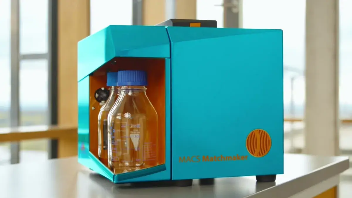
How scientists use MACS Matchmaker
Crude samples
Pre-purification eats weeks. Your binder fails when the matrix changes from buffer to lysate or serum.
Measure directly in serum, lysate, or cell supernatant. No purification, no method redevelopment.
Background masks signal
Labels and NSB hide the real binding event. Real hits slip through as false negatives.
Focal molography reads coherent mass density. Non-specific binding and matrix noise stay structurally invisible.
Multi-week panels
Sequential SPR sweeps stall every decision. Cross-species FcRn, biosimilar comparability, off-rate ranking all wait in line.
Eight pre-conjugated ligands on one chip. The full cross-species panel runs in approximately one hour.
Sub-twofold gaps
Critical 1.5× differences disappear. Below the NSB floor, the data you need to rank candidates is invisible.
Eight within-chip replicates per ligand. Confidence intervals from a single run resolve sub-twofold differences.
One pair at a time
Conventional kinetics runs sample-by-sample. 64 simultaneous interactions stay on the to-do list.
Up to 64 simultaneous interactions on a single chip. One injection series delivers the full panel.
Drift-limited weak binders
Slow off-rates wash away in baseline drift before a clean curve can be fit.
Drift below 0.05 pg/mm²/min. Long association phases stay rock-stable, even at low-µM K_D.