


The MACS® Matchmaker
Every target deserves its perfect match.
Experience a robust platform that delivers quantitative data on binder affinity, kinetics, and specificity in real samples.
The MACS® Matchmaker measures binding kinetics directly in crude media, without labels or purification. It is built for drug discovery and interaction studies that buffer-only assays cannot answer.
- Label-free measurements in crude media: Measure under in vivo-like conditions and eliminate pre-purification, saving time.
- Simple assay development: Use our easy-to-use sensor functionalization protocol to set up experiments faster.
- Multiplexing for increased speed: Characterize 64 interactions simultaneously to significantly reduce experiment times.
- Assay automation: Use the MACS® Sampler for fully unattended runs with up to 2 × 384-well plates.
- Drift-minimized data: Obtain robust, reliable data with fewer artifacts through our patented focal molography technology.
- pM sensitivity: Characterize even small proteins or peptides in complex biological matrices.
- Remote instrument control and enhanced connectivity: Control and access the instrument from any device at any time.
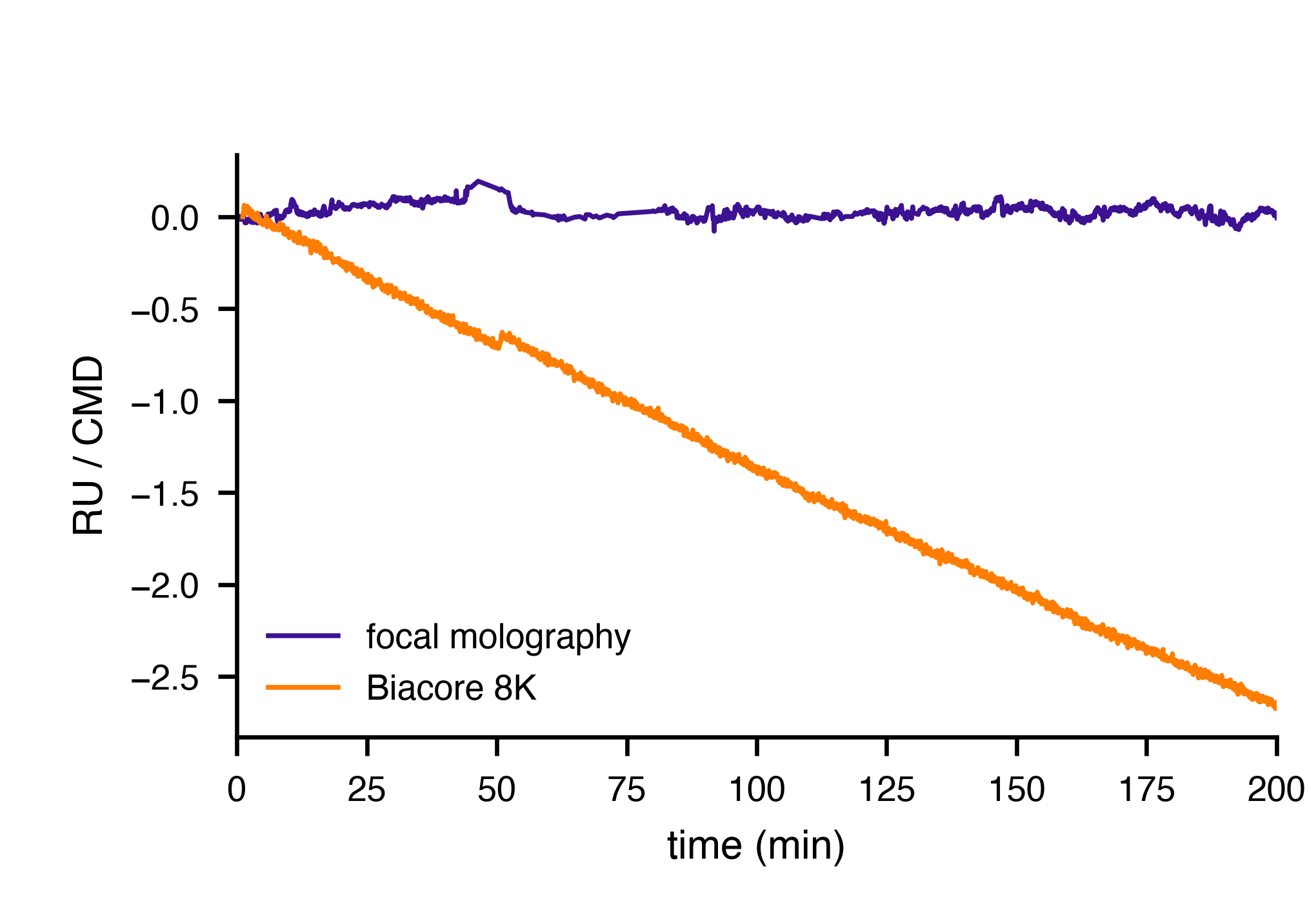
Drift-minimized baseline
Baseline traces for focal molography and a Biacore 8K channel over more than three hours under running buffer flow. No binding signal change is expected.
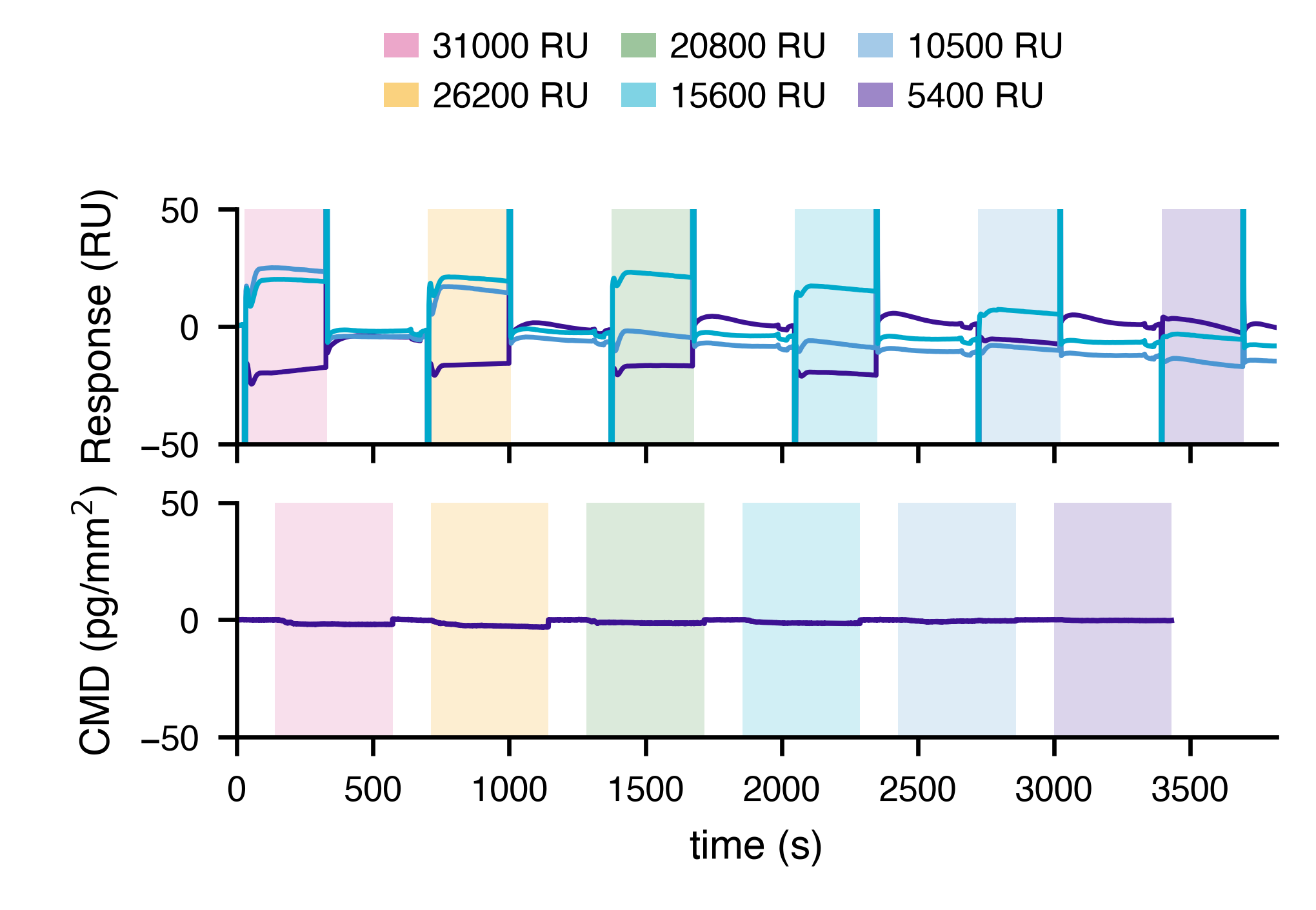
Lowest medium influence on the market
Molography has no artefacts from buffer changes during the measurement. The tested SPR setup showed stronger responses to buffer changes, even after referencing. [Experiment was the injection of different glycerol concentrations; no binding signal is expected]
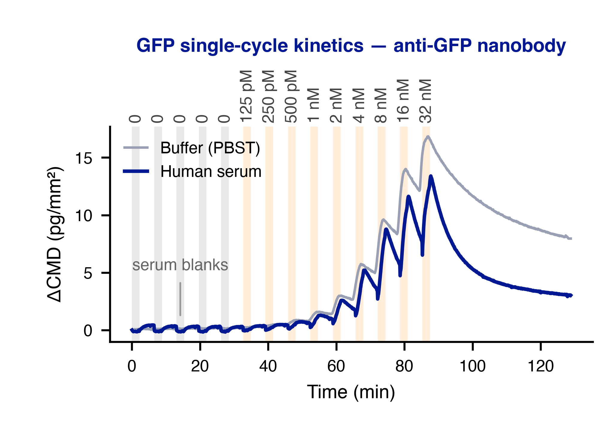
Makes crude samples look like buffer
GFP single-cycle kinetics on an anti-GFP nanobody in human serum, compared with PBST buffer. Serum blanks are followed by a GFP titration from 125 pM to 32 nM.
The MACS® Matchmaker is powered by focal molography.
Focal molography is a biophysical technique that enables robust and sensitive detection of biomolecular interactions without the need for fluorescent labels. This method facilitates the analysis of these interactions within complex biological samples, such as serum, bioreactor fluids, or cell culture media.
The technique operates by diffracting laser light through a specialized two-dimensional nanopattern of molecular binding sites on a sensor chip, known as a mologram. This mologram functions as a focusing diffractive lens, directing light into a precise focal point. The intensity of this focused light correlates with the quantity of molecules bound to the mologram, thereby indicating the extent of biomolecular interactions.
A significant advantage of focal molography is its resilience against environmental noise, such as temperature fluctuations and non-specific binding of off-target molecules. This robustness eliminates the need for temperature stabilization or sensor equilibration, making it a versatile tool for various applications.
Overall, focal molography expands the analytical capabilities for studying biomolecular interactions across a wide range of biological research and diagnostic applications.
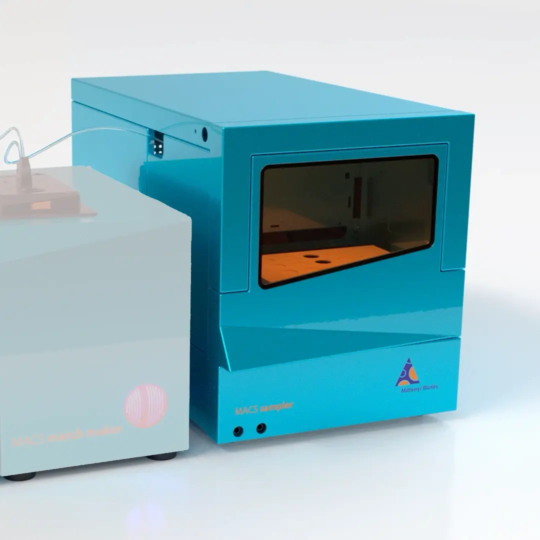
Simplify your workflow and save valuable laboratory time with the fully automated MACS® Sampler.
Depending on your needs, you can combine the MACS® Matchmaker with the MACS® Sampler, a fully integrated automation solution for interaction studies.
- Sample format: 300 µL or 2 mL vials; 96- or 384-well plates.
- Capacity: Two positions for 48-vial trays or well plates.
- Throughput: Characterize up to 1,536 interactions per day.*
- Flow path: One needle and one syringe pump.
- Hands-off operation: Up to 72 h of unattended run time.
- Biocompatibility: Metal-free sample flow path with a biocompatible sample needle, valves, and tubing.
- Cooling: The sample compartment can be chilled to 4 °C for optimal storage.
- Software: Direct integration with the MACS® Matchmaker software.
*With DNA-directed immobilization (DDI) chemistry in batches of 64 immobilized compounds against one target.
The specifications. At a glance.
Explore binding performance, imaging, sample handling and data analysis.