Heterogeneous vs. Homogeneous Assays
After giving examples for practical examples of Biosensors, todays articles should classify differences between heterogeneous and homogeneous assays. To properly understand the differences between both assays, a few definitions need to be clarified: Assay: testing procedure for estimating the concentration of a pharmaceutically active substance in a formulated product or bulk material (1).
Antibody: specialized Y-shaped proteins produced by our immune system that bind like a lock-and-key to the body’s foreign invaders — whether they are viruses, bacteria, fungi or parasites (3). When antibodies bind their specific targets, our immune cells recognize the invader covered in antibodies more efficiently, and can then eliminate it.
Antigen: any substance, living or not, that triggers an immune response.
What is the difference?
Both of these assay classes most commonly regroup what we call immunoassays, “bioanalytical methods that measure the presence or concentration of analytes in a solution through the use of an antibody or an antigen as a biorecognition agent.”(1) Homogeneous assays can yield accurate read-outs without having to separate the analyte of interest from the biomolecules (e.g. labeled antibodies) used to detect it. On the other hand, heterogeneous assays require one or more steps of separation, where unbound antibodies and/or unbound analyte must be washed away. This makes heterogeneous assays often longer and more complex to conduct; however, they are often more precise than homogeneous assays, and can be very helpful to detect more complex analytes. Homogeneous assays are usually reserved for the detection of small, simple molecules.
EMIT- A Homogeneous Assay
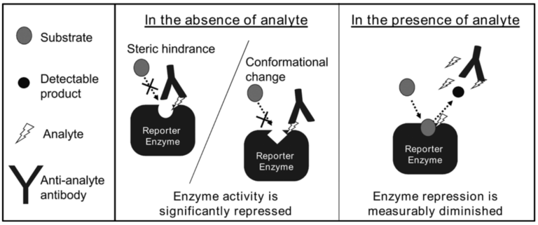
Enzyme Multiplied Immunoassay Technique, otherwise known as EMIT, is an assay used to detect the presence or the amount of analyte in a solution. As its name implies, it uses enzymes to yield this result. The main component of the assay is a complex made of an enzyme (“reporter enzyme” in figure below), attached to the analyte of interest. The enzyme’s active site (the part that is specific for binding) is specific for a substrate that is fluorescent after its binding event with the enzyme. In short, when this substrate and the enzyme-analyte complex bind, the substrate changes color (yielding a detectable product). These complexes are added to a sample solution, usually issued from a patient; when the color change of the substrate occurs, the whole sample solution will change color. This however still does not help us explain how we can determine the presence of the analyte of interest in the sample. To this end, we add an antibody specific for the analyte of interest to the sample (anti-analyte antibody). Here, there are two scenarios: either the sample contains the analyte of interest, or it does not.
If the sample does not contain the analyte of interest, the antibody will only be able to bind it whilst attached to the enzyme (in the enzyme-analyte complex), because there is no free analyte in the present in the sample solution. When this happens, the antibody blocks the active site of the enzyme. This means that no other molecule can bind this site, because there is no access possible. The color-substrate that would usually bind there and become fluorescent can therefore no longer bind; it never becomes fluorescent and there is no color change in the sample. If, however, the solution does contain the analyte of interest, the added antibodies have many more molecules to bind: they will primarily bind the free analyte in solution (because of its easy access), as well as some of the enzyme-analyte complexes. Nevertheless, some of these complexes will remain unbound by the antibody; there the color-substrate can bind and become fluorescent; the sample then changes color as well.
Mechanism of EMIT
For a more animated explanation, go watch the video at : EMIT immunoassay
ELISA- A Heterogeneous Assay
ELISA stands for Enzyme Linked Immunosorbent Assay; like its homogeneous counterpart EMIT, it is a heterogeneous assay that can determine the presence or the quantity of an analyte.
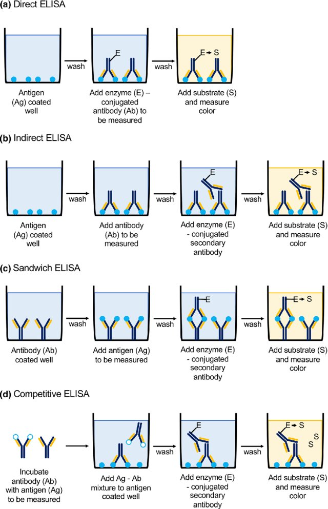
There are four types of ELISA assays: indirect, sandwich, and competitive ELISA (see figure below). All three involve an analyte, at least one antibody, and an enzyme that changes color when a desired reaction occurs. What makes all types of ELISA heterogeneous is the washing steps (also depicted), that eliminate any non-bound molecules, be it the analyte of interest or their specific antibody.
One of the most commonly used ELISA is the indirect assay. Typically, it is used to detect the presence of an antigen, for example a virus. The test begins by attaching the antigen to the bottom of a plate. After washing the plate to remove any unbound antigen, we add an antibody specific for this antigen (primary antibody). After another wash step to remove any unbound antibodies, we add another antibody (secondary antibody), this one specific for the first antibody. The secondary antibody is usually issued from an animal (e.g. rabbit), so that it recognizes the human antibody administered first. It is also conjugated to an enzyme that will react with a substrate that we administer in the final step of the assay. Before we add this substrate, we wash the full sample one more time to remove any unbound secondary antibody. Finally, we add the substrate: when reacting with the enzyme, a color change is induced, and the solution changes color. The color change indicates the presence of the analyte of interest, and the color change is proportional to the amount of primary antibody present, and thereby also to the amount of analyte.
Comparison between various ELISA assays
As shown in the figure on the left, there are other types of ELISA; each provides various advantages and disadvantages, displayed in the table on the right.
Did you know? Immunoassays and SARS-CoV-2
One of the biggest disadvantages of ELISA in the past was its low throughput readout. Roche has simultaneously solved this problem and created a fast detection method of SARS-Cov-2 based on their general Elecsys assay technique. It is “an immunoassay for the in vitro qualitative detection of antibodies (including IgG) to SARS-CoV-2 in human serum and plasma”. It utilizes a principle very similar to ELISA called ECLIA, pictured below.
References
- Biological Assays, Introduction to Biological Assays
- LiveScience, What are antibodies?
- Ju, H., Lai, G. and Yan, F., 2017. Immunosensing for detection of protein biomarkers. Elsevier.
- SlideShare, Homogeneous and heterogeneous immunoassay
- Engvall, E., 1980. [28] Enzyme immunoassay ELISA and EMIT. In Methods in enzymology (Vol. 70, pp. 419-439). Academic Press.
- Abcam, ELISA: basic principles and types of ELISA assay
- Elecsys® Anti-SARS-CoV-2
Images
- Pharmaceutical Analysis, Chapter 24 – Page 8
- Boguszewska, K., Szewczuk, M., Urbaniak, S. and Karwowski, B.T., 2019. immunoassays in DNA damage and instability detection. Cellular and Molecular Life Sciences, pp.1-16.
- Elecsys® Anti-SARS-CoV-2
- Chiu, M.L., Lai, D. and Monbouquette, H.G., 2011. An influenza hemagglutinin A peptide assay based on the enzyme-multiplied immunoassay technique. Journal of Immunoassay and Immunochemistry, 32(1), pp.1-17.
