


This recording is available on demand
Register with a short form to watch the full recording.
Access the recordingAbout the talk
Recorded at the 3rd focal Molography User Meeting on September 9, 2026. Szabolcs Novák (HUN-REN Centre for Energy Research) shows how focal molography follows the adhesion of living cancer cells to RGD-functionalized sensor chips and separates where integrins bind from how much mass arrives at the surface.
Topics covered
- From molecules to living cells. Integrins link the cell to its surroundings, recognize the RGD peptide motif and are overexpressed in many tumors. The Nanobiosensorics Laboratory has studied their adhesion kinetics with label-free biosensors, single-cell force methods and holographic microscopy, but evanescent-field sensors cannot tell specific integrin binding from nonspecific mass.
- An open-channel cell protocol. HeLa cells are pipetted onto click chemistry chips that carry RGD on the mologram ridges and a protein-repellent PEG layer in the grooves, then imaged by microscopy. Vinculin staining confirms that the cells adhere through integrins on the molograms.
- Three phases of adhesion. In the first phase, coherent and total mass rise together as nascent adhesions form on the ridges. In the second phase, the coherent signal falls while mass keeps arriving, because focal adhesions grow out into the grooves. In the third phase, the coherent signal recovers as adhesions mature back along the ridges.
- Integrin numbers per cell. With an analyte efficiency of about 10%, the integrin count per HeLa cell lands in the same order of magnitude as earlier estimates from other biosensors, which supports this value as a working approximation.
- Cells respond to treatment. EDTA releases the cells, neuraminidase removes sialic acid from the glycocalyx and gives a weaker, slower coherent signal, and histamine changes adhesion in line with earlier measurements. A first experiment on DNA chips with cyclic RGD points to reusable surfaces.
More videos and webinars
 14:19
Sep 9, 2026
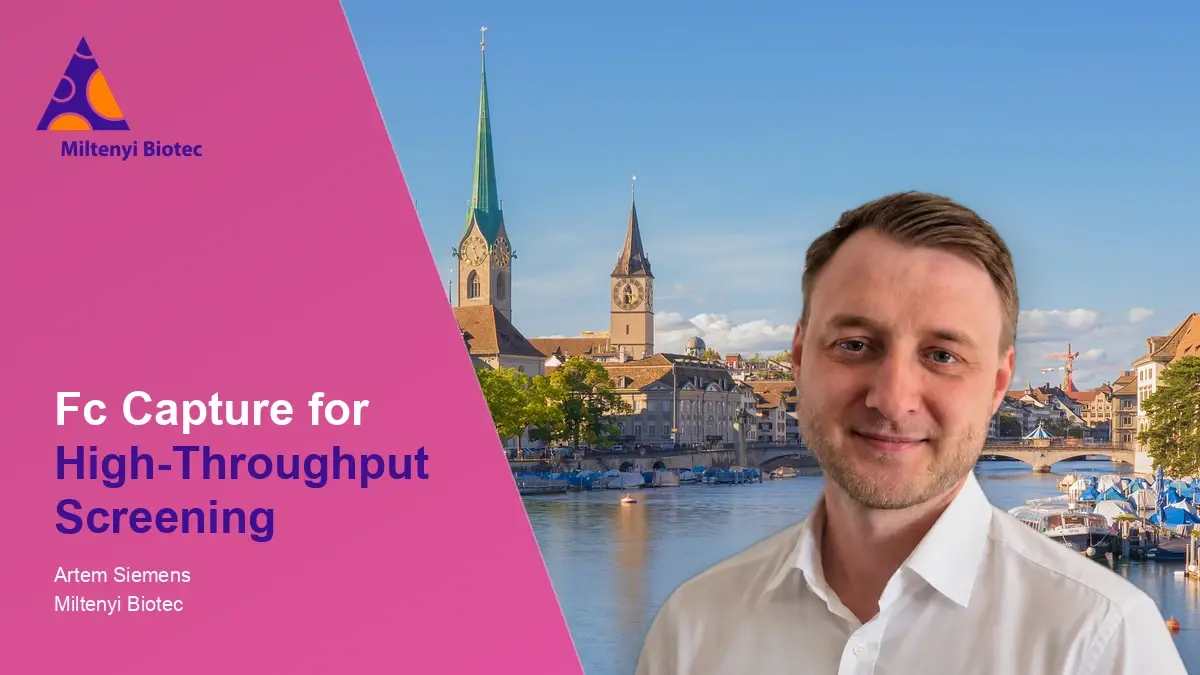
Development of an Fc-Capturing Protein for High-Throughput Screening at Miltenyi Biotec
Artem Siemens
Recorded at the 3rd focal Molography User Meeting, Artem Siemens (Miltenyi Biotec) shows how a homodimeric capture nanobody, immobilized by DNA-directed immobilization (DDI), ...
Watch after a short form
 31:56
Sep 9, 2026
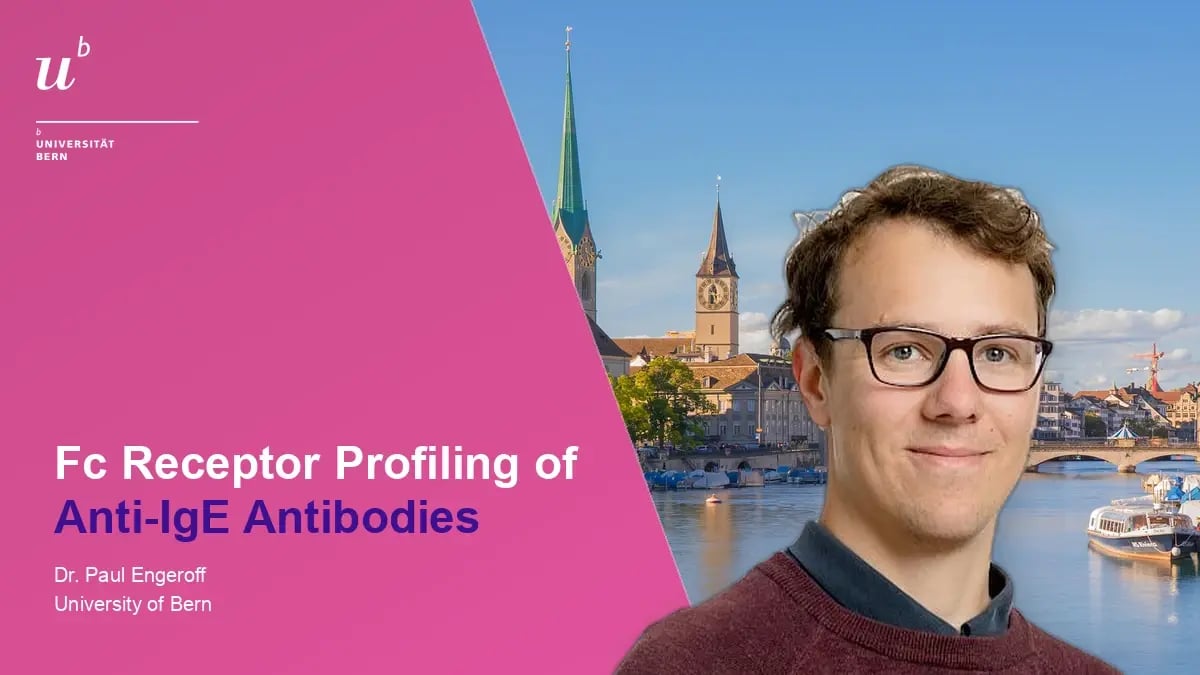
Focal molography for the Fc receptor profiling of therapeutic anti-IgE antibodies
Dr. Paul Engeroff
A multiplex Fc receptor panel on focal molography maps where omalizumab, ligalizumab and their immune complexes with IgE and allergen bind, across all human Fc gamma and IgE ...
Watch after a short form
 26:18
Sep 9, 2026
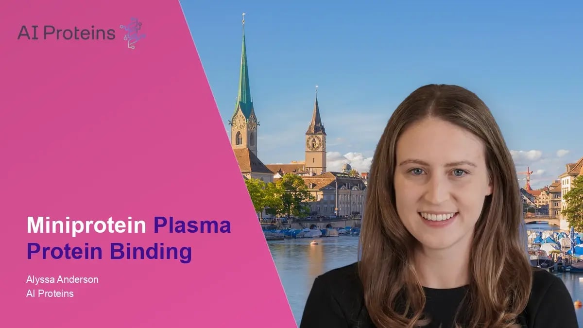
Focal Molography Enables Rapid Evaluation of Miniprotein Plasma Protein Binding
Alyssa Anderson
Plasma protein binding of 15 designed miniproteins measured directly in plasma on one 64-plex chip, validated against ultracentrifugation and extended by kinetic information.
Watch after a short form