


Our earlier articles covered common examples of label-based technologies (see Practical Examples of Biosensors). This article discusses a widely used label-free technology: surface plasmon resonance (SPR).
What is SPR?
Surface plasmon resonance (SPR) is an optical biosensing technology that detects molecular interactions. The name describes both the technology and the physical effect behind it. SPR has been in use for about 35 years and detects analytes in solution with very high sensitivity.
How does it work?
The SPR effect occurs when transverse magnetic (TM) polarized light hits the interface between a metal and a dielectric material such as glass. Light that travels from a medium with a higher refractive index (n1) into one with a lower index (n2) can be totally reflected. The figure below shows this. It happens at any angle θ above a threshold angle that can be calculated.
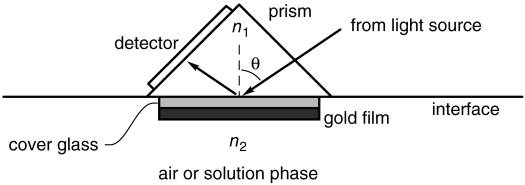
How can we detect and quantify molecular interactions with this setup?
We start with a gold film and cover it with a protein coating that repels unwanted molecules. Into this coating we embed ligands that recognize the analyte of interest (recognition molecules). The coated gold film sits on a glass layer, coating facing up. The sample that may or may not contain the analyte (mobile phase) flows over this surface. If the analyte is present, it binds to its ligands. When polarized light hits the surface at a specific angle θ, a new wave is excited: the surface plasmon (SP) mode. Forming this wave takes energy, so part of the energy that would go into the reflected light goes into the plasmon instead. At the angle θ where the plasmon forms, almost all the light energy goes into the SP mode and the reflected light is nearly extinguished.
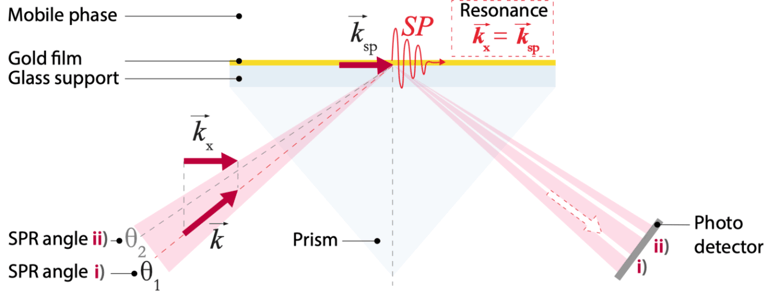
When the SPR biosensor is prepared, the angle θ at which this effect occurs is recorded. This angle matters because it shifts slightly whenever a molecule binds to the recognition molecules on the gold film. Binding changes the refractive index close to the surface, and that in turn changes the angle at which the plasmon is launched.
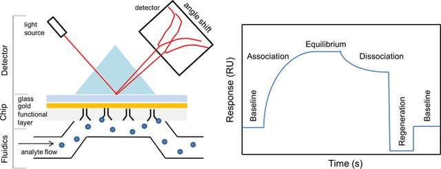
While the sample flows over the coated gold film, a photodetector array measures the reflected light intensity as a function of angle (figure below). This intensity profile depends on the refractive index close to the surface, which depends on the amount of bound analyte. The angle shift therefore tells us how much analyte binds to the sensor surface. Plotted over time, this gives a binding curve, the sensorgram (see figure 3).
For an animated explanation of SPR, visit https://youtu.be/o8d46ueAwXI.
Hungry for more?
Are you wondering how sensorgrams differ between samples with and without analyte, or between analytes with high and low affinity? Head over to Surface plasmon resonance | Cytiva, formerly GE Healthcare Life Sciences, for animated sensorgrams in these conditions.
Advantages and disadvantages of SPR
SPR has advantages over many other biosensing technologies because it gives sensitive, real-time, and quantitative information. Its sensitivity is high: it detects down to a picogram of molecules per mm2. That is about 1/300 of a monolayer of water molecules on a surface. Because the measurement runs in real time, it yields the affinity and the kinetics of the interaction, which drug development depends on. The sensorgram carries this information; it is drawn in real time while the binding reaction runs (see example below). Real-time measurements are valuable in biological research, even more so when they are label-free (see the article “What is a biosensor”), as with SPR. Because SPR is so sensitive, a few microliters of sample are enough.
SPR also has its limits. The main one is that SPR measures only the change in refractive index at the sensor surface; it is an integrative sensor. Think of a bathroom scale: put apples and oranges on it and you get one total weight. The scale cannot tell the apples from the oranges or count them. Temperature, changes of medium, and non-specific binding also change the refractive index at the surface. Non-specific binding means molecules that bind the surface but that we do not want to detect. As with the scale, we do not know exactly what we are reading. SPR reacts strongly to these outside influences and needs stabilization. It also needs long equilibration times, buffers cannot be switched during a measurement, and interactions can only be measured in well-defined buffers. Real biological samples contain many background molecules that also bind to the sensor surface (non-specific binding).
These non-specific interactions have lower affinity than the specific interaction with the analyte of interest. But in most samples there are far more non-specific molecules than specific ones, often a million to a billion times more. Their binding therefore hides the specific signal and limits detection. This cross-sensitivity of SPR to medium changes, temperature, and non-specific binding is its environmental noise problem. On top of that, the angle shifts that binding causes are tiny. The angle has to be measured with a relative precision of 10-5, which is like seeing the Eiffel Tower from China. That takes expensive scientific cameras and equipment.
Did you know?
SPR is a well-proven technology for analyzing molecular interactions, but it reaches its limits in crude biological samples and in measurements that are not stabilized. The angle shift of the SPR resonance also has to be measured with extreme precision.
Why is that? Why are SPR measurements so demanding? Could a technology keep the advantages of SPR (label-free, real-time, sensitive, quantitative) and also work in non-purified samples? Could it filter out environmental and experimental noise? Our next article answers this question.
References
Tang, Y., Zeng, X. and Liang, J., 2010. Surface plasmon resonance: an introduction to a surface spectroscopy technique. Journal of chemical education, 87(7), pp.742-746.
Surface plasmon resonance | Cytiva, formerly GE Healthcare Life Sciences
Images
Katharina Kübrich, 2019. Molographic Peptide Arrays: Towards Label-Free Protein Signaturing in Undiluted Blood Plasma. ETH Zürich Master’s Thesis, p. 12.
Tang, Y., Zeng, X. and Liang, J., 2010. Surface plasmon resonance: an introduction to a surface spectroscopy technique. Journal of chemical education, 87(7), pp.742-746.
Damborský, P., Švitel, J. and Katrlík, J., 2016. Optical biosensors. Essays in biochemistry, 60(1), pp.91-100