


 By
·
7 minute read
Fc receptors are the docking sites that let immune cells grab onto antibodies. Small differences between people's receptors change how tightly an antibody binds. Whether that matters for patients is harder to answer, and it depends on the antibody, the disease, and the treatment. Here is what the evidence shows, and what it means for how you characterize your next antibody candidate.

If you develop antibodies, the variants of the Fcγ receptors (FcγR) belong in your characterization. These variants are called allotypes. Testing a candidate against each variant shows how it binds, and it tells you which functional experiments to run next.
Two receptors, two variants each
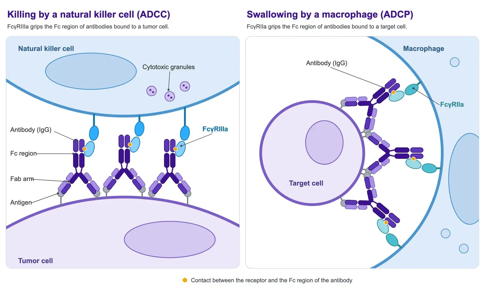
An antibody has two jobs. Its arms bind the target. Its tail, the Fc region, calls in immune cells by docking onto Fcγ receptors on their surface.
People carry slightly different versions of these receptors. Two of the best-studied examples are FcγRIIIa (versions V158 and F158) and FcγRIIa (versions H131 and R131).
- FcγRIIIa sits on natural killer cells and some other immune cells. It helps them kill cells that carry antibodies, a process called antibody-dependent cellular cytotoxicity (ADCC). The V158 version generally binds human IgG1 antibodies more strongly than the F158 version.
- FcγRIIa helps immune cells to react and to swallow antibody-coated particles. This process is called antibody-dependent cellular phagocytosis (ADCP). The H131 and R131 versions differ most clearly in how they bind IgG2, another antibody type.1
These two receptors are a good starting point for asking how receptor binding relates to antibody function.
An early signal in lymphoma
The first clinical signal came from lymphoma. In 2002, Cartron and colleagues studied 49 patients with follicular lymphoma, a type of blood cancer, who had not been treated before. Each patient received rituximab alone. Rituximab is an antibody that marks cancer cells so that immune cells can destroy them.
The researchers then checked which version of FcγRIIIa each patient carried. FCGR3A is the gene for this receptor. Patients with two copies of the V version (called 158V homozygous) responded better than patients who carried an F version.
After two months, 100% of the V/V patients had responded, against 67% of the others. After twelve months, the numbers were 90% and 51%. Both differences had a p-value of 0.03, which means chance alone is an unlikely explanation.2
The antibody was the same in both groups. Only the receptor differed. So the strength of antibody binding became a possible explanation, and a property worth measuring.
A small study of cetuximab then showed that the story is less simple. Cetuximab is an antibody used against colorectal cancer. The study followed 39 patients whose cancer had spread. The patients received cetuximab alone.
The FcγRIIa version (gene name FCGR2A) was linked to progression-free survival, the time before the disease gets worse. For FcγRIIIa, the link was only a trend (p = 0.055), just short of the usual threshold of 0.05.
Patients with the best combination of both receptor versions stayed free of progression for a median of 3.7 months. The median is the middle value. For the other patients, it was 1.1 months.
Curiously, the better FCGR3A group included patients with an F version. Yet V158 is the version that generally binds IgG1 more strongly.3
Then the picture got more complicated
Larger studies did not consistently reproduce these links:
- An international consortium collected samples from 1,123 patients with metastatic colorectal cancer. They were treated with cetuximab alone or with chemotherapy. In patients with no KRAS mutation in the tumor (KRAS wild-type), two groups were checked. These were FCGR2A-HH (two H copies) and FCGR3A-VV (two V copies). Neither group had significantly longer progression-free survival.4
- In the RESORT study, the receptor genes of 321 patients were read. These patients had follicular lymphoma with a low tumor burden, meaning only a small amount of tumor. No FCGR2A or FCGR3A genotype predicted the first response to rituximab alone.5
The picture also differs between diseases. A later study looked at 835 patients with rheumatoid arthritis or systemic lupus erythematosus, two autoimmune diseases. It found links between the FCGR3A-158V version and clinical or biological responses to rituximab.6
In short: Larger studies did not reproduce the early links, and the results differ between diseases. Whether a receptor version matters depends on the setting. Receptor binding works together with other factors. These include how much target is on the cell and how many immune cells are available. They also include how much drug reaches the tissue, and other ways the treatment acts. A difference in binding strength alone cannot tell you how an individual patient will respond.
What a controlled experiment can show
In the laboratory, under defined conditions, the link between receptor version and function becomes clearer. Dall'Ozzo and colleagues compared how well natural killer cells from VV donors and FF donors killed target cells with rituximab:7
- At very high rituximab concentrations, both groups killed equally well.
- The concentration needed to kill half of the target cells differed 4.2-fold: 0.00096 µg/mL for VV donors and 0.00402 µg/mL for FF donors (p = 0.0043).
- The difference appeared only at concentrations that barely marked the CD20-positive target cells. CD20 is the protein that rituximab binds.
In short: In the laboratory, a receptor version can change how much antibody is needed for the same effect. This may contribute to differences between clinical settings, but it does not prove their cause. Whether the same shift matters at low tissue exposure or low target density has to be tested in the relevant system.
From allotype to drug design: margetuximab
In the SOPHIA trial, about 86% of patients carried at least one F158 copy9. So the weaker-binding version of FcγRIIIa (also called CD16A) is the common one.
Margetuximab is an antibody against HER2, a protein found on some breast cancer cells. Its Fc region is engineered to bind both CD16A versions more strongly than unmodified IgG1. The idea behind this: in several studies of trastuzumab, another anti-HER2 antibody, patients with an F version responded less well. These patients might benefit from a stronger call to the immune system.8
The phase 3 SOPHIA trial compared margetuximab with trastuzumab, each combined with chemotherapy. It included 536 patients with HER2-positive advanced breast cancer who had been treated before.
The results use two measures. HR is the hazard ratio: a value below 1 means a lower risk that the disease gets worse or that the patient dies. CI is the 95% confidence interval, the range in which the true value most likely lies.
- Progression-free survival improved with margetuximab: median 5.8 versus 4.9 months (HR 0.76; 95% CI 0.59–0.98).8
- Overall survival did not improve: median 21.6 versus 21.9 months in the final analysis (HR 0.95; 95% CI 0.77–1.17).9
- By CD16A genotype, exploratory analyses pointed in different directions. In 158FF patients (two F copies), overall survival was possibly better with margetuximab: median 23.6 versus 19.2 months (HR 0.72; 95% CI 0.52–1.00). In 158VV patients (two V copies), it was possibly better with trastuzumab: median 31.1 versus 22.0 months (HR 1.77; 95% CI 1.01–3.12).9
The trial authors note that there is no clear biological explanation for the lack of benefit in the VV group.8 The genotype analyses were planned in advance, but they are exploratory and need confirmation.
Even so, they show that stronger binding to both versions does not guarantee the same outcome in each genotype group. This is why the binding profile of a candidate deserves a close look before it reaches the clinic.
What this means for your candidate
For a candidate in development, the practical question is how its receptor-binding profile relates to the activity your program wants to achieve. A useful characterization strategy includes:
- Both common versions of FcγRIIa and FcγRIIIa. Compare H131 with R131, and V158 with F158, under matched assay conditions.
- The broader receptor profile. Include other relevant activating receptors and the inhibitory receptor FcγRIIb. This shows how selective an engineered or natural Fc region is.
- A suitable reference molecule. Measure the candidate next to an established comparator of the same class and format.
- Relevant functional assays. Use ADCC, ADCP, or other assays that fit the mechanism of action. They show how binding differences affect cellular responses.
A note on the numbers: equilibrium affinity (KD) tells you how tightly two molecules bind. It is one part of the profile. The association rate constant (kon) says how fast they bind. The dissociation rate constant (koff) says how fast they let go. Both add information where the interaction and the assay allow reliable kinetic resolution.
Multivalent immune complexes, which join several antibodies together, add a further effect. Their apparent binding also depends on avidity, the combined grip of several binding sites, and on the assay geometry. So define the molecular state and the experimental setup.
Measuring the profile with the Fc Receptor Panel
The Fc Receptor Panel on the MACS Matchmaker measures receptor binding without labels, using focal molography. DNA-directed immobilization (DDI) attaches the receptors to the chip. This allows many receptors to be measured side by side, and the panel can be configured as needed.
A possible panel includes FcγRIIa and FcγRIIIa. Both receptors can be measured in both versions (H131 and R131, V158 and F158), so a study can compare the versions side by side. The panel composition can be adapted to your question.
These measurements give a development team a direct way to compare candidates, find changes that affect only one receptor, and choose the next functional experiment. Doing this early gives the program a stronger basis for interpreting later biological results.
References
- Bruhns, P. et al. (2009) Specificity and affinity of human Fcγ receptors and their polymorphic variants for human IgG subclasses. Blood 113: 3716–3725. doi:10.1182/blood-2008-09-179754
- Cartron, G. et al. (2002) Therapeutic activity of humanized anti-CD20 monoclonal antibody and polymorphism in IgG Fc receptor FcγRIIIa gene. Blood 99: 754–758. doi:10.1182/blood.v99.3.754
- Zhang, W. et al. (2007) FCGR2A and FCGR3A polymorphisms associated with clinical outcome of epidermal growth factor receptor expressing metastatic colorectal cancer patients treated with single-agent cetuximab. J. Clin. Oncol. 25: 3712–3718. doi:10.1200/JCO.2006.08.8021
- Geva, R. et al. (2015) FCGR polymorphisms and cetuximab efficacy in chemorefractory metastatic colorectal cancer: an international consortium study. Gut 64: 921–928. doi:10.1136/gutjnl-2014-307234
- Kenkre, V. P. et al. (2016) Fc gamma receptor 3A and 2A polymorphisms do not predict response to rituximab in follicular lymphoma. Clin. Cancer Res. 22: 821–826. doi:10.1158/1078-0432.CCR-15-1848
- Robinson, J. I. et al. (2022) Comprehensive genetic and functional analyses of Fc gamma receptors influence on response to rituximab therapy for autoimmunity. EBioMedicine 86: 104343. doi:10.1016/j.ebiom.2022.104343
- Dall'Ozzo, S. et al. (2004) Rituximab-dependent cytotoxicity by natural killer cells: influence of FCGR3A polymorphism on the concentration-effect relationship. Cancer Res. 64: 4664–4669. doi:10.1158/0008-5472.CAN-03-2862
- Rugo, H. S. et al. (2021) Efficacy of margetuximab vs trastuzumab in patients with pretreated ERBB2-positive advanced breast cancer: a phase 3 randomized clinical trial. JAMA Oncol. 7: 573–584. doi:10.1001/jamaoncol.2020.7932
- Rugo, H. S. et al. (2023) Margetuximab versus trastuzumab in patients with previously treated HER2-positive advanced breast cancer (SOPHIA): final overall survival results from a randomized phase 3 trial. J. Clin. Oncol. 41: 198–205. doi:10.1200/JCO.21.02937