


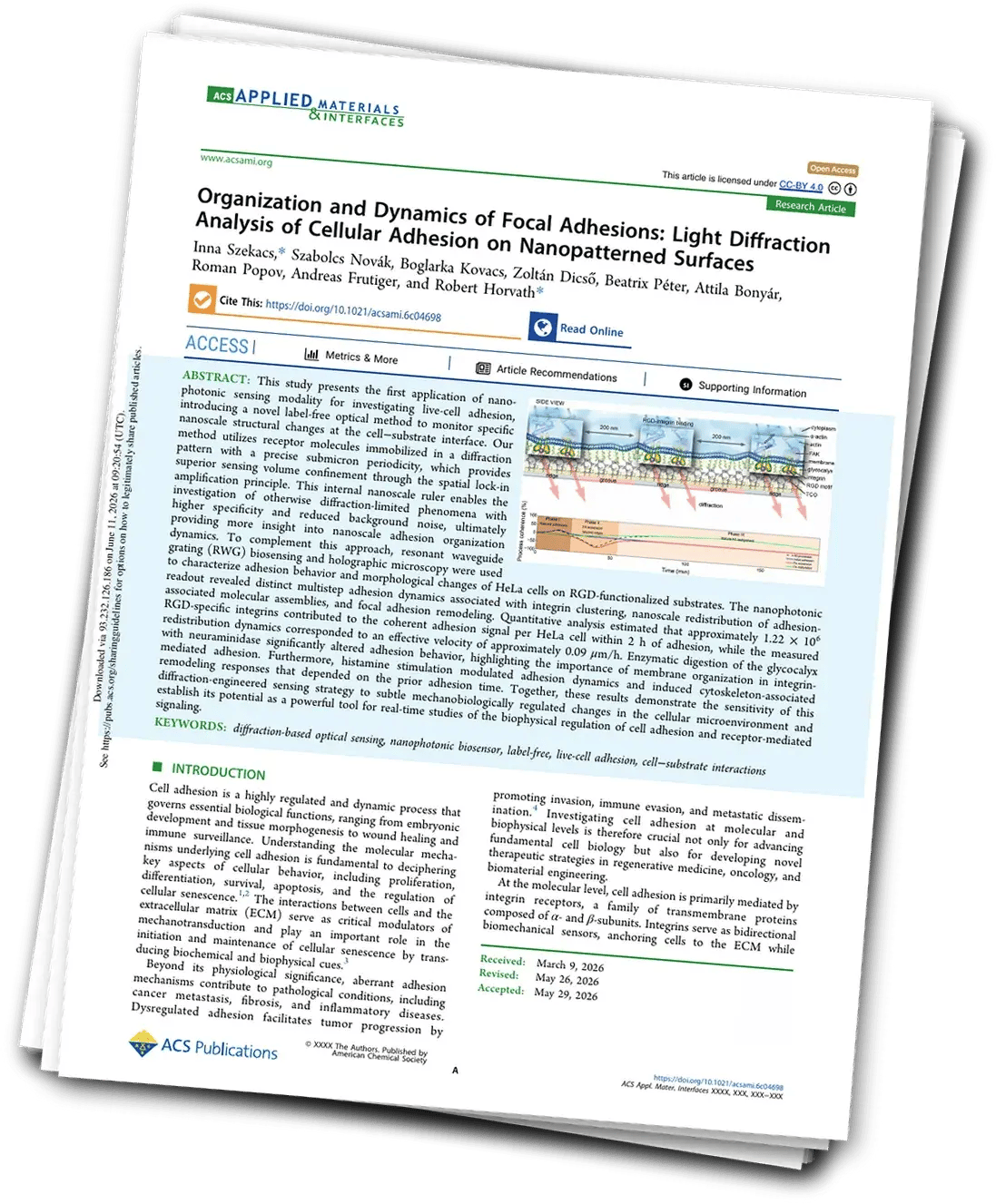
Organization and Dynamics of Focal Adhesions: Light Diffraction Analysis of Cellular Adhesion on Nanopatterned Surfaces
Focal molography resolves live-cell adhesion into three distinct phases, quantifying over one million integrins per HeLa cell in real time, label-free.
Get the publicationThe Challenge
Cell adhesion governs proliferation, differentiation, immune surveillance and metastasis. Yet biologists studying cell-substrate interactions face a tradeoff. Fluorescent labels can perturb the very process they want to watch. Traditional label-free biosensors like resonant waveguide grating (RWG) cannot separate specific receptor engagement from bulk refractive-index drift or non-specific membrane contact.
The Approach
The team functionalized MACS® Matchmaker sensor chips with sub-micron RGD patterns (200 nm ridges, 200 nm grooves) using bioorthogonal TCO-tetrazine click chemistry. Then they seeded HeLa cells onto the surface. Focal molography selectively detects molecules that occupy the engineered pattern. The recorded signal therefore comes from integrin engagement at RGD sites, not from glycocalyx, membrane proteins, or cytosolic background.
Key Results
- Three adhesion phases resolved: Phase I (0–23 min), early integrin engagement on RGD ridges. Phase II (23–54 min), lateral expansion of adhesion assemblies beyond ridges. Phase III (54–192 min), mature focal-adhesion alignment along stress-fiber axes.
- 1.22 × 10⁶ RGD-specific integrins per HeLa cell quantified at 2 h adhesion, consistent with literature on integrin density.
- 0.09 μm/h redistribution velocity of integrin-associated adhesion assemblies between ridges and grooves, derived from sinusoidal pattern modulation.
- Glycocalyx contribution measured: neuraminidase digestion shifted adhesion kinetics, demonstrating sensitivity to membrane-level remodeling.
- Histamine modulation state-dependent: stimulation effects scaled with the maturation state of focal adhesions at the time of stimulus.
Why It Matters
Focal molography opens a label-free, quantitative window into mechanobiology, integrin signaling, and focal-adhesion dynamics on engineered substrates. The method is directly relevant for cancer-adhesion research, regenerative medicine, and drug discovery against mechanotransduction targets. Thanks to focal molography's high specificity in complex matrices, it could extend to whole-tissue samples.
Szekacs, I., Novák, S., Kovacs, B., Dicső, Z., Péter, B., Bonyár, A., Popov, R., Frutiger, A., & Horvath, R. (2026). Organization and Dynamics of Focal Adhesions: Light Diffraction Analysis of Cellular Adhesion on Nanopatterned Surfaces. ACS Applied Materials & Interfaces. https://doi.org/10.1021/acsami.6c04698
More from our resource library you may like
 Oct 2026
High-throughput thermodynamic fingerprinting of protein–ligand interactions by DNA-directed focal molography
Oehninger, J., Notova, S., & Frutiger, A. (2026). PLOS One, 21(10), e0354008. https://doi.org/10.1371/journal.pone.0354008
Download after a short form
 Oct 2026
Same target, different stickiness: polyreactivity of therapeutic antibodies
Six clinical antibodies, including two same-target pairs, ranked by polyreactivity against an eight-ligand panel on one focal molography chip. Briakinumab scored 69-fold above ...
Download after a short form
 Oct 2026
One on-DNA ligand against many targets: selectivity profiling for DEL hit validation
On-DNA oncoFAP profiled against human, mouse and canine FAP and five unrelated proteins on one focal molography chip layout. Full kinetics on every species variant, flat responses ...
Download after a short form